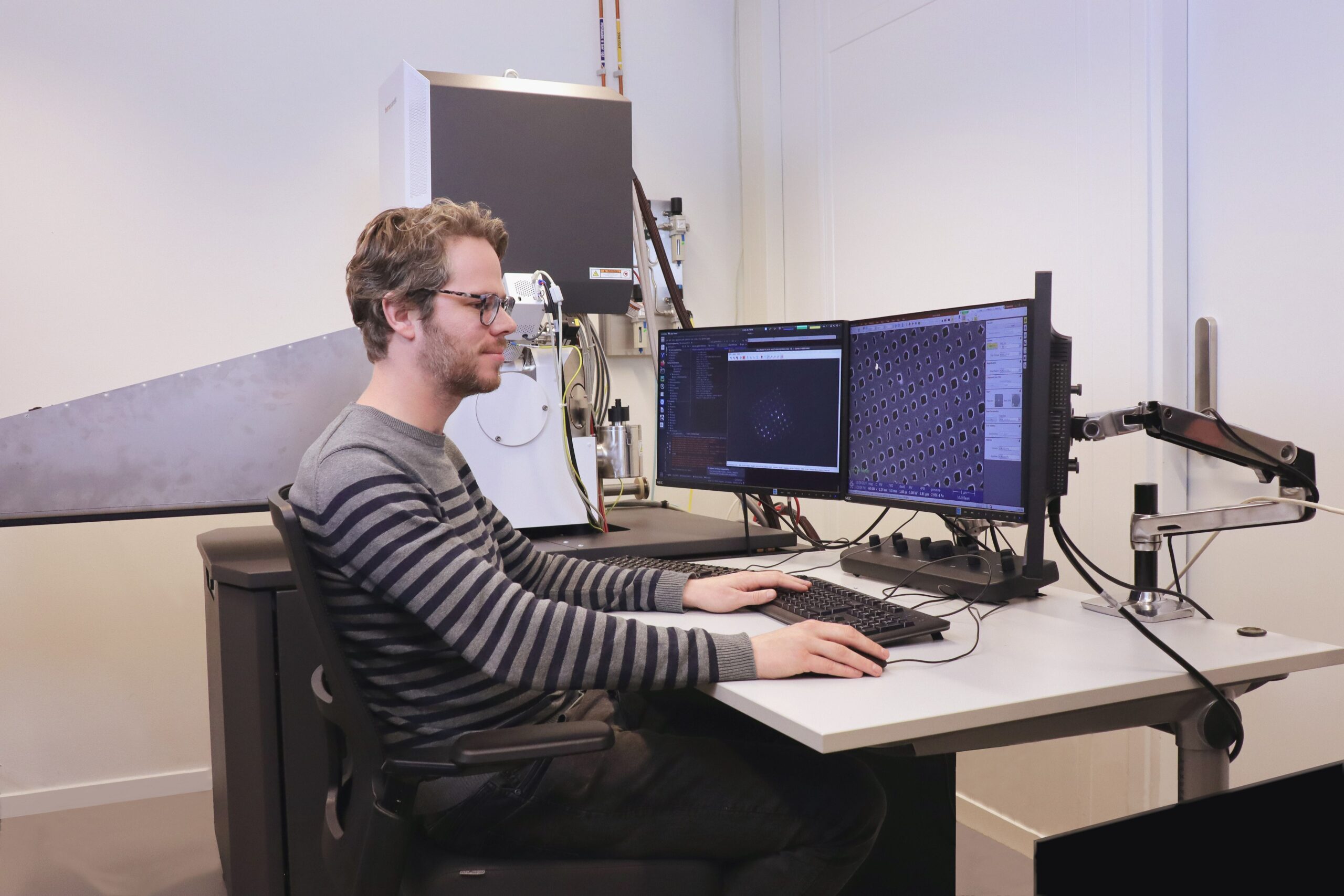
FAST-EM High-speed electronics for a multibeam scanning electron microscope
TNL is currently working together in a consortium with – amongst others – Delft University of Technology and Delmic to develop an innovative electron microscope, a so-called Scanning Electron Miscroscope (SEM). SEMs can make a preparation visible by irradiating it with an electron beam. By analyzing the electrons that are scattered back and the electrons emitted by the material, a SEM can visualize preparations to within a nanometer. Just to refresh your memory: a nanometer is one billionth of a meter, about as large as five atoms in a row.
High-speed technology for data transport
Scanning with a single-electron-beam SEM takes a lot of time. It takes hours to fully scan a preparation with a surface of one square millimeter. But the scanning time can be strongly reduced by scanning simultaneously with multiple electron beams. The consortium is therefore developing the FAST-EM: an SEM system with multiple electron beams, or – in short – a multibeam SEM. Scanning with multiple electron beams generates very large volumes of data. Within the project, TNL is realizing very high-speed electronics for data transport and detection.
Image processing and analysis
One of the challenges of multibeam electron microscopy is that the scans generated by each individual electron beam subsequently have to be assembled. Each beam scans a rectangular piece of the sample. Because the electron beams are guided by a magnetic lens, these scanned rectangles never match each other exactly. This means the scans must always overlap each other by a few pixels. Calibration can help to determine where the scans made by the various beams match up. An additional challenge for scans of biological samples is to generate sufficient contrast. Therefore we are developing algorithms for imaging and analysis to create accurate images of the samples that are of sufficient quality for medical and scientific purposes.
Further development
Delmic’s FAST-EM has now been implemented succesfully by early adopters, such as the University Medical Center Groningen and Delft University of Technology. The speed of the scanner offers new possibilities for research. The UMCG is using the FAST-EM to realize an atlas of the human body, which allows easy retrieval of characteristic properties and prominent elements of medical samples. The Delft University of Technology utilizes the FAST-EM for brain research and the research into the pathogenesis of brain diseases.
The consortium will continue the further development of the FAST-EM. Besides TNL data platform and other projects, Delmic is developing a cutting robot for automated preparation and scanning of the thousands of samples that are required for doing detailed 3D scans.